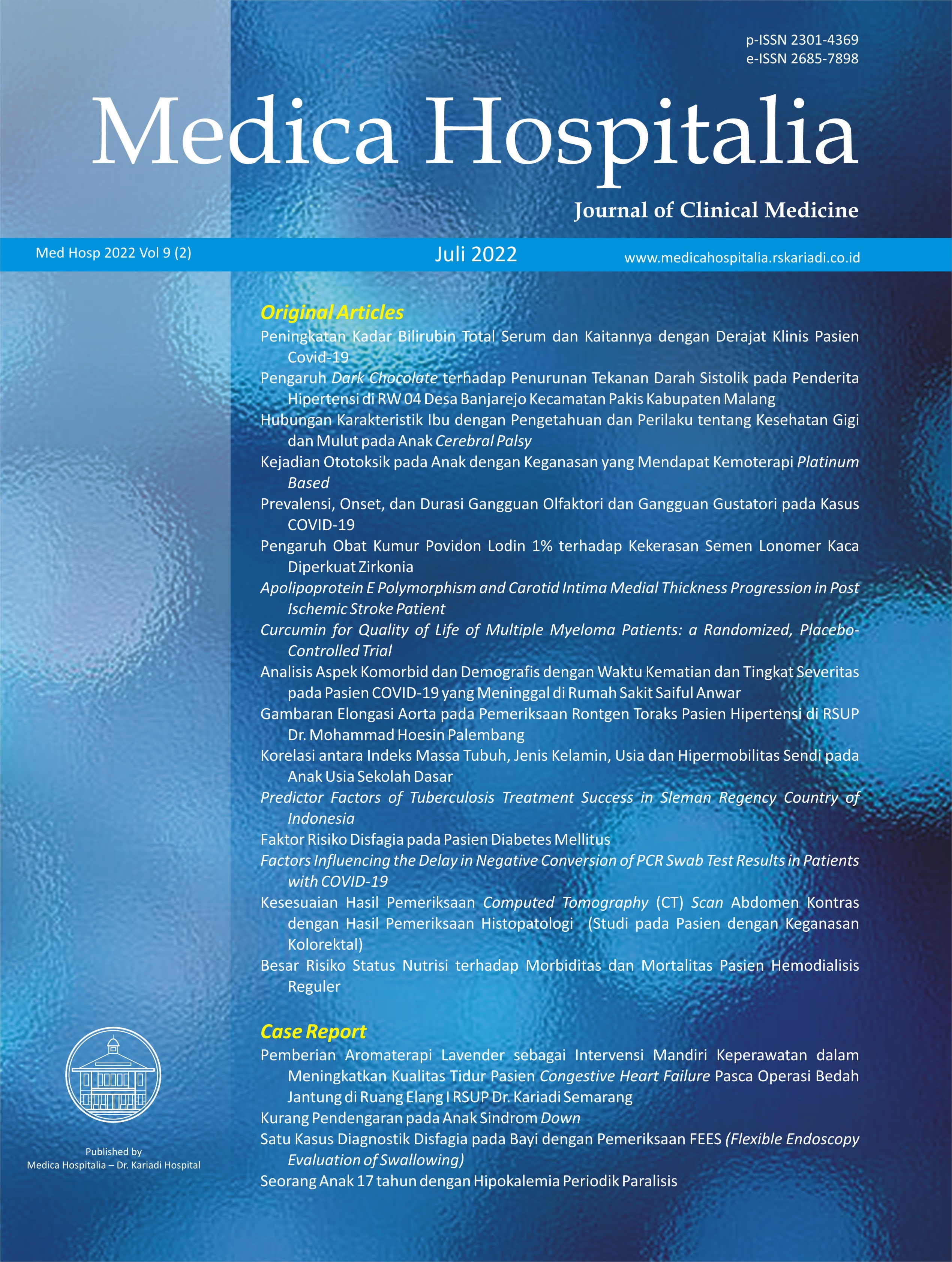
Kesesuaian Hasil Pemeriksaan Computed Tomography (CT) Scan Abdomen Kontras dengan Hasil Pemeriksaan Histopatologi (Studi pada Pasien dengan Keganasan Kolorektal)
Suitability Computed Tomography (CT) Scan Abdomen Contrast Results with Histopathological Examination Results (Studies in patients with colorectal malignancies)
DOI:
https://doi.org/10.36408/mhjcm.v9i2.760Keywords:
CT Scan, Histopatologi, Keganasan kolorektal, StagingAbstract
Latar belakang : CT Scan abdomen kontras adalah modalitas pencitraan yang sering digunakan pada pasien dengan kecurigaan keganasan kolorektal seperti adenokarsinoma, neuroendocrine tumor (NET), gastrointestinal stromal tumor (GIST) dan limfoma karena mampu menskrining, mendiagnosis sekaligus menilai staging. Ketepatan diagnosis dan staging akan berpengaruh terhadap tatalaksana selanjutnya. Penelitian ini bertujuan untuk mengetahui kesesuaian hasil pemeriksaan CT Scan abdomen kontras dengan hasil pemeriksaan histopatologi mengenai karakteristik, jenis dan staging lokal pada pasien dengan keganasan kolorektal.
Metode : Penelitian ini merupakan studi observasional dengan pendekatan cross- sectional. Terdapat 61 subyek penelitian yang dilakukan penilaian karakteristik, jenis dan stagingnya menggunakan CT Scan oleh dua ahli radiologi konsultan abdomen sedangkan pemeriksaan histopatologi dilakukan oleh ahli patologi anatomi konsultan abdomen. Uji diagnostik dan uji kesesuaian dilakukan untuk menganalisis kesesuaian hasil pemeriksan CT Scan dan histopatologi.
Hasil : Berdasarkan karakteristik pada CT Scan, 100% sampel termasuk keganasan yang mengarah pada jenis karsinoma, sehingga kesesuaian karakteristik dan jenis tidak dapat dilakukan. Adapun untuk staging (CT Scan) didapatkan T3 57,4% dan T4 42,6%. Pada pemeriksaan histopatologi didapatkan 95,1% adenokarsinoma, 3,3% GIST dan 1,6% limfoma dengan staging pT3 65,6% dan pT4 34,4%. Didapatkan konsistensi dalam penilaian staging lokal antara pemeriksaan CT Scan abdomen kontras dan pemeriksaan histopatologi dengan nilai sensitivitas 82,5%, spesifisitas 90%, nilai prediksi positif 94%, nilai prediksi negatif 73%, tingkat akurasi 85% serta nilai kappa 0,691.
Simpulan : CT Scan abdomen kontras dapat digunakan sebagai modalitas pencitraan untuk staging pada pasien keganasan kolorektal dengan konsistensi cukup baik.
Downloads
References
Rawla P, Sunkara T, Barsouk A. Epidemiology of colorectal cancer: Incidence, mortality, survival, and risk factors. Prz Gastroenterol. 2019;14(2):89–103.
Kementrian Kesehatan Republik Indonesia/Kemenkes RI. Laporan Nasional Riset Kesehatan Dasar 2018 [Internet]. Badan Penelitian dan Pengembangan Kesehatan. 2018. p. 674. Available from: http://labdata.litbang.kemkes.go.id/images/ download/laporan/RKD/2018/Laporan_Nasional_RKD201 8_FINAL.pdf
Karacin C, Türker S, Eren T, Imamoglu GI, Yılmaz K, Coskun Y, et al. Predictors of Neoplasia in Colonic Wall Thickening Detected via Computerized Tomography. Cureus. 2020;12(9).
Richie AJ, Mellonie P, Suresh HB. Diagnostic Accuracy of Pre- operative Staging of Colorectal Carcinoma in Comparison to Postoperative Pathological Staging. Int J Sci Study. 2016;4(4):38–41.
So JS, Cheong C, Oh SY, Lee JH, Kim YB, Suh KW. Accuracy of preoperative local staging of primary colorectal cancer by using computed tomography: Reappraisal based on data collected at a highly organized cancer center. Ann Coloproctol. 2017;33(5):192–6.
Elibol FD, Obuz F, Sökmen S, Terzi C, Canda AE, Sağol Ö, et al. The role of multidetector CT in local staging and evaluation of retroperitoneal surgical margin involvement in colon cancer. Diagnostic Interv Radiol. 2016;22(1):5–12.
Dahlan MS. Statistik untuk Kedokteran dan Kesehatan: Deskriptif, Bivariat, dan Multivariat, Dilengkapi AAplikasi dengan Menggunakan SPSS. 2013. 159 p.
Levy AD, Sobin LH. From the archives of the AFIP - Gastrointestinal carcinoids: Imaging features with clinicopathologic comparison. Radiographics. 2007; 27(1):237–57.
Yoshida T, Kamimura K, Hosaka K, Doumori K, Oka H, Sato A, et al. Colorectal neuroendocrine carcinoma: A case report and review of the literature. World J Clin Cases. 2019;7(14):1865–75.
Vernuccio F, Taibbi A, Picone D, La Grutta L, Midiri M, Lagalla R, et al. Imaging of gastrointestinal stromal tumors: From diagnosis to evaluation of therapeutic response. Anticancer Res. 2016;36(6):2639–48.
Reddy RM, Fleshman JW. Colorectal gastrointestinal stromal tumors: A brief review. Clin Colon Rectal Surg. 2006;19(2):69–77.
Gay ND, Chen A, Okada CY. Colorectal Lymphoma: A Review. Clin Colon Rectal Surg. 2018;31(5):309–16.
Singla S, Kaushal D, Sagoo H, Calton N. Comparative analysis of colorectal carcinoma staging using operative, histopathology and computed tomography findings. Int J Appl Basic Med Res. 2017;7(1):10.
Sultana N, Khan S, Baloch S. Diagnostic accuracy of contrast enhanced computed tomography in staging of colorectal carcinoma. Pakistan Armed Forces Med J. 2018;68(5):1076–81.
Zhou XC, Chen QL, Huang CQ, Liao HL, Ren CY, He QS. The clinical application value of multi-slice spiral CT enhanced scans combined with multiplanar reformations images in preoperative T staging of rectal cancer. Med (United States). 2019;98(28).
Korsbakke K, Dahlbäck C, Karlsson N, Zackrisson S, Buchwald
P. Tumor and nodal staging of colon cancer: accuracy of preoperative computed tomography at a Swedish high-volume center. Acta Radiol Open. 2019;8(12):205846011988871.
Thornton E, Mendiratta-Lala M, Siewert B, Eisenberg RL. Patterns of fat stranding. Am J Roentgenol. 2011;197(1):1–14.
Pereira JM, Sirlin CB, Pinto PS, Jeffrey RB, Stella DL, Casola E G. Disproportionate fat stranding: A helpful CT sign in patients with acute abdominal pain. Radiographics. 2004;24(3):703–15.
Additional Files
Published
How to Cite
Issue
Section
Citation Check
License
Copyright (c) 2022 Muhammad Beni, Maya Nuriya Widyasari, Devia Eka Listiana, Titik Yuliastuti

This work is licensed under a Creative Commons Attribution-ShareAlike 4.0 International License.
Copyrights Notice
Copyrights:
Researchers publishing manuscrips at Medica Hospitalis: Journal of Clinical Medicine agree with regulations as follow:
Copyrights of each article belong to researchers, and it is likewise the patent rights
Researchers admit that Medica Hospitalia: Journal of Clinical Medicine has the right of first publication
Researchers may submit manuscripts separately, manage non exclusive distribution of published manuscripts into other versions (such as: being sent to researchers’ institutional repository, publication in the books, etc), admitting that manuscripts have been firstly published at Medica Hospitalia: Journal of Clinical Medicine
License:
Medica Hospitalia: Journal of Clinical Medicine is disseminated based on provisions of Creative Common Attribution-Share Alike 4.0 Internasional It allows individuals to duplicate and disseminate manuscripts in any formats, to alter, compose and make derivatives of manuscripts for any purpose. You are not allowed to use manuscripts for commercial purposes. You should properly acknowledge, reference links, and state that alterations have been made. You can do so in proper ways, but it does not hint that the licensors support you or your usage.


